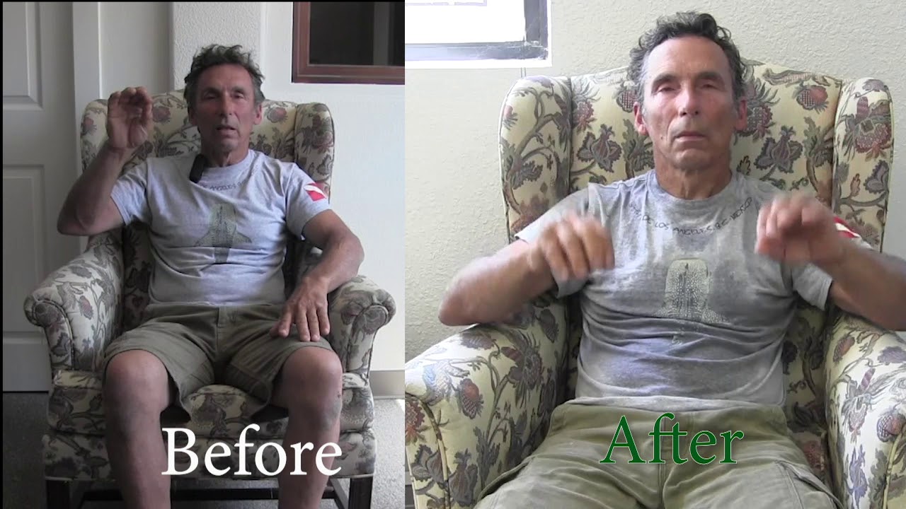
Before and After: What 2 Training Sessions Did to Parkinson’s Motor Function
A subject with a 5+ year Parkinson’s diagnosis completed two LiveO2 sessions. The before and after tests — finger tap, heel-toe, balance — show something most neurologists don’t expect to see.
The Experiment Setup
This case study records motion performance in an individual who has lived with a Parkinson’s-related motor diagnosis for over five years. The subject completed two LiveO2 Adaptive Contrast sessions and was tested before and after the second session.
One key detail: before testing was conducted while the subject was generally medicated. After testing was done while generally unmedicated. This makes the observed improvements more notable, not less — the post-session results were achieved without the chemical support present in the baseline.
Test Protocol
Motor Tests Used
- Finger tap test (bilateral)
- Lightbulb movement (wrist rotation)
- Heel and toe tap
- Standing balance
Conditions
- Before: 2nd session, generally medicated
- After: post-session, generally unmedicated
- 5+ year neurological pathology
- Single subject case study
What the Camera Recorded
Motor Function Changes
Finger Tap Test
Before: one hand noticeably slower, hard to sustain the rhythm, described as “working with a nut and bolt — sometimes hard to put that nut on the bolt.” After: more sustained, less effortful, both hands more consistent.
Lightbulb Movement (Wrist Rotation)
Before: right hand harder than left, requiring noticeable effort. After: reduced resistance, smoother rotation. Subject noted the effort differential between hands decreased.
Heel and Toe Tap
Before: right side better than left; freeze events present. After: movement more fluid, freeze events reduced. “Sometimes it almost freezes — that’s what they call the freeze.”
Standing Balance
Before: legs felt disconnected, eyes-open balance compromised. After: more stable stance, improved eyes-closed test performance. “I look like a normal human.”
“It’s very subtle feeling. Hard to describe. It’s kinda like working with this nut and bolt — sometimes it would be hard to put that nut on the bolt.”
— Subject describing the difference in hand function during testingWhy Oxygen Changes Motor Function
Motor neurons fire electrochemical signals to produce movement. That process consumes oxygen. When oxygen supply to motor regions is compromised — whether from vascular dysfunction, disease progression, or simple altitude — the signals degrade. Movements become slower, less precise, and more likely to freeze.
Parkinson’s pathology is primarily about dopamine-producing neurons in the substantia nigra. But a significant secondary factor is vascular — reduced blood flow to motor control regions compounds the dopaminergic loss.
What Oxygen Training Does
LiveO2 Adaptive Contrast alternates between oxygen-rich and oxygen-reduced breathing during cardiovascular exercise. This drives large volumes of oxygenated blood through the circulatory system, including to the brain and nervous system.
When motor neurons receive more oxygen, they fire more reliably. The freeze events that characterize Parkinson’s motor dysfunction have a real-time oxygen component — and when that component is addressed, function improves measurably.
This is not a cure or reversal of the disease. It is a functional improvement driven by better oxygen availability — and it requires ongoing maintenance, like any exercise intervention.