Measuring Brain Oxygenation from LiveO2
You can’t improve what you can’t measure. Here’s how we prove the brain gets more oxygen.
Watch: Brain Oxygenation Testing After LiveO2
See real-time fNIRS data showing what happens to brain oxygen after a session.
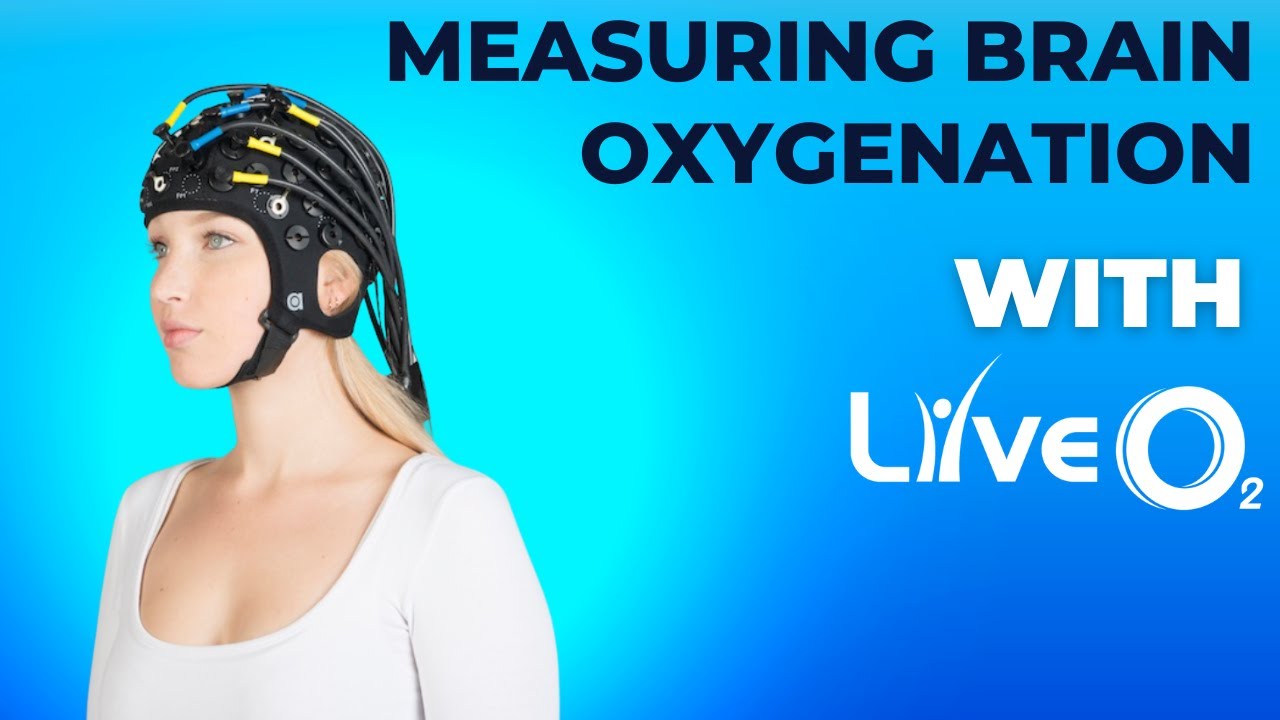
In this clip, we review how brain oxygenation is tested after training with LiveO2 using functional near-infrared spectroscopy (fNIRS) — a medical-grade tool that reads oxygen levels in brain tissue through the skull.
What Is fNIRS?
fNIRS stands for functional near-infrared spectroscopy. It works by shining safe infrared light through the skull. That light bounces off your blood. Oxygenated blood and deoxygenated blood absorb light differently. The sensor reads the difference.
That gives you a live map of how much oxygen is reaching your brain tissue — not just your lungs, not just your finger. Your actual brain.
Hospitals and research labs use fNIRS to study cerebral oxygenation during exercise. It’s the same technology used in stroke units and brain injury clinics. It’s non-invasive, portable, and gives real-time data.
“We use the same fNIRS equipment that brain researchers use in clinical trials. It shows exactly what happens to brain oxygen before and after a LiveO2 session.”
— LiveO2 Research TeamWhy Measuring Brain Oxygen Matters
Most people guess how well their brain is doing. They feel foggy or sharp. Tired or alert. But feelings are not data.
When you measure brain oxygenation, you get a number. Before the session. After the session. The difference tells you exactly what changed.
This is the same approach used in a 2012 study on fNIRS and cognitive function — researchers tracked how oxygen levels in the prefrontal cortex changed during mental tasks. Higher oxygen meant better performance.
More oxygen to the brain = better thinking. fNIRS lets you see that in real time — not weeks later, not based on a feeling. Right now.
That’s the difference between hope and proof. LiveO2 doesn’t ask you to take our word for it. We show you the data.
What Happens After a LiveO2 Session
Here’s the pattern we see over and over with fNIRS readings:
Before the session: Brain oxygenation sits at a baseline. For most people — especially those over 50 or dealing with brain fog — that baseline is lower than it should be. Narrowed capillaries, poor circulation, and inflammation all reduce how much oxygen reaches brain tissue.
During the session: Adaptive Contrast drives blood flow up. You breathe low oxygen to trigger vasodilation. Your heart pushes harder. Then you switch to high oxygen. Your lungs flood that fast-moving blood with dissolved oxygen.
After the session: fNIRS shows a measurable increase in brain tissue oxygenation. Users report feeling clearer, sharper, and more focused — and the data backs it up.
The whole process takes 15 minutes. The measurement takes seconds. The results speak for themselves.
Who Benefits from Brain Oxygen Testing
People with brain fog. If you feel like you’re thinking through mud, your brain is likely low on oxygen. Measuring confirms it — and shows the improvement after training. Read more about brain fog and oxygen.
Concussion and TBI recovery. Brain injuries damage the tiny blood vessels that carry oxygen. fNIRS shows whether oxygen delivery has been restored. Learn about concussion and oxygen.
Older adults. As we age, capillaries narrow. Brain oxygen drops. Testing gives you a number to track over time — and proof that training is working.
High performers. Athletes and executives who want to know their brain is running at full capacity. One test before your session. One test after. The data tells the story.
Anyone who wants proof, not promises. LiveO2 isn’t about guessing. It’s about measuring.
Common Questions
LiveO2 uses fNIRS (functional near-infrared spectroscopy) devices from Artinis. These are medical-grade sensors that shine infrared light through the skull to read oxygen levels in brain tissue. The same technology is used in hospitals and research labs worldwide.
Yes. fNIRS uses near-infrared light — the same kind your TV remote uses. It’s completely non-invasive. No radiation. No needles. No discomfort. You just wear a lightweight sensor on your forehead.
The change is immediate. fNIRS readings taken right after a 15-minute LiveO2 session show measurable increases in brain tissue oxygenation. Most users also feel the difference — clearer thinking, better mood, more energy.
LiveO2 offers before-and-after neurological panels so you can see your own data. Many users take cognitive assessments alongside fNIRS readings to track both the oxygen delivery and the performance improvement.
Yes. A pulse oximeter clips onto your finger and measures oxygen saturation in your blood. fNIRS measures oxygen in your brain tissue directly. Your blood can be 98% saturated and your brain can still be starved — because the issue is delivery, not supply. fNIRS shows whether oxygen is actually getting where it needs to go.
Yes. The BrainO2 protocol is designed specifically for brain oxygenation. It uses the Adaptive Contrast challenge to drive maximum blood flow to the brain, then floods it with high-concentration oxygen.